|
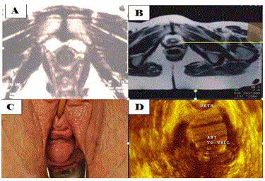
| Figure 5 Images, by MRI, 3DUS and a clinical image of the vagina and the IUS. Image A is a cross section by MRI, the IUS is a compact collagen-muscle tissue cylinder, and the vagina in a nullipara is an H-shape. Image B is a MRI image, cross section, in a patient who suffers from SUI and vaginal prolapse; the IUS is torn with an open lumen. The vagina is lax, stretched and prolapsed. Image D is a 3DUS image, which shows the anterior vaginal wall, prolapsed. Image C is the clinical picture of vaginal prolapse. |